by Lodi Vet | May 3, 2017 | Cats, Dogs, Educational Materials, Wellness
Parasites, or worms, are organisms that live at the expense of other animals. Dogs and cats commonly host parasitic infections which may be a health concern for both you and your pet. Intestinal parasites live primarily in the stomach and intestines where they feed off the host tissues and intestinal content. The amount of damage done depends on the type and number of parasites. Intestinal parasites are diagnosed by checking a fresh stool sample for eggs which are shed by the adult worm in the intestinal tract.
There are four species of worms commonly seen in dogs and cats:
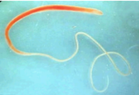
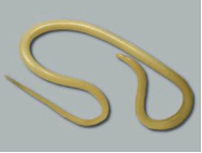 ROUNDWORMS
These are longer (2-4 inches), round, white worms which may be seen in vomit or passed in the stool. Puppies and kittens may be infected by the mother while in the uterus or while nursing (through the milk). Older pets are infected by contact with contaminated stool/soil or eating other animals (rabbits, mice, rats & earthworms) that have been exposed to roundworm eggs. In large numbers, these worms may cause malnutrition, diarrhea, pot-bellied appearance and intestinal obstruction.
 HOOKWORMS
These worms are microscopic (not seen with the naked eye) and can be the most harmful of the internal parasites. The mouth parts of this worm have a number of hooks which tear at the intestinal wall causing excessive blood loss. Infection occurs through contact with contaminated stool/soil. Hookworm larvae can actually penetrate the skin, or puppies may be infected through the mother’s milk. Bloody diarrhea, weight loss, and anemia are possible signs of a severe hookworm infection.
 WHIPWORMS
Whipworms are also microscopic worms which live in the lower intestinal tract. They can be difficult to diagnose because the eggs are shed intermittently. Infection occurs by eating the eggs which can remain in the environment for a long time.
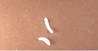 TAPEWORMS
Tapeworm segments are shed intermittently and can be seen as small, rice-like segments either in the stool or attached to the hair around the anus. Fecal exams may not pick up this parasite. Animals become infected by eating rabbits, birds and rodents or through fleas. They do not directly get tapeworms from other animal’s stool.
PUBLIC HEALTH SIGNIFICANCE:
- Human infection with roundworm and hookworm larvae is possible but does not occur frequently.
- Eating contaminated stool or soil (roundworms) or direct contact with infected soil (hookworms) is necessary for human infection.
- Children are at greatest risk because of their play and indiscriminate eating habits.
INTESTINAL PARASITE PREVENTION:
Have a routine fecal exam checked every 6-12 months.
- Fresh samples less than 12hrs old. Refrigerate if > 2hrs. Avoid samples that have been in contact with soil for prolonged times.
- Clearly identify the stool donor (avoid sample mix-ups)
- Kitty litter mixed with sample is acceptable as long as the sample is fresh.
- Avoid frozen samples or dried out, hard samples.
- Collect in a clean, sealed container. (Provided upon request)
Once diagnosed use a specific dewormer for the type of parasite found.
- Follow the recommended deworming schedule and recheck fecals as directed.
- Over the counter, broad-spectrum dewormers are often not effective.
Do not allow animals to urinate/defecate in children’s play area or sand box.
Prevent your pet from eating rabbits, rodents, birds and control fleas (tapeworms).
Check fecal and deworm dogs before breeding and again prior to whelping to prevent/reduce infecting puppies.
Deworm puppies at 3 weeks and 6 weeks of age. Follow up with a fecal exam at 12 weeks of age.
- Eggs can remain infective in soil for years, especially in dog pens or areas where pets are tied.
- Replace dirt runs with concrete.
- Turn soil over to depth of 8-12 inches after pet has been dewormed.
- Move your pet to a new uncontaminated area.
- Remove feces from your lawn or kennel daily
Practice and teach children good hygiene when playing with animals, especially puppies and kittens.
Walk and exercise your dog in areas not frequented by other dogs. Avoid public parks, playgrounds, hunting grounds etc..
Use a monthly heartworm preventative that also helps control intestinal parasites. We recommend using Interceptor Plus as your monthly heartworm preventative and broad-sprectrum intestinal parasite preventative.
by Lodi Vet | May 3, 2017 | Cats, Dogs, Educational Materials
Pancreatitis is a condition caused by inflammation of the pancreas, an organ of the digestive tract. The pancreas is located in the abdomen, lying close to the stomach and first part of the intestines. Functions of the pancreas include controlling blood sugar by producing a hormone called insulin and producing enzymes responsible for the digestion of fats and proteins.
Although the healthy pancreas produces useful enzymes, these same enzymes can cause the pancreas to destroy its own tissue. This process called “auto-digestion” is the first step in the development of pancreatitis. In mild cases with supportive care, changes in diet, and short courses of medications, the pet will recover. In severe cases, the tissues of the pancreas become damaged or cells of the pancreas begin to die off (necrosis), causing enzymes from the pancreas to leak into the abdomen resulting in pain and inflammation. Bacteria can then invade this area causing fluids to accumulate in the pancreas and abdomen leading to shock and possible death.
The causes of pancreatitis can be numerous, however in 90% of the cases the exact cause is unknown, or idiopathic. Sometimes it is due to overeating or eating high fat foods. Other predisposing causes include trauma to the pancreas, cancer, or inflammatory bowel disease.
Symptoms of pancreatitis include vomiting, lack of appetite, diarrhea, lethargy, restlessness and discomfort in the abdomen.
Diagnosis is by clinical signs and blood testing. Ultrasound is a helpful diagnostic tool to image the pancreas and surrounding areas to look for signs of other problems like tumors, fluid leakage or inflammation of the intestines. Early diagnosis and treatment of pancreatitis will give your pet the best chance of recovery.
Treatment of pancreatitis depends on the severity. Mild cases of pancreatitis can be treated with oral medications and bland diet. More severe cases can require hospitalization, intravenous fluids and medications. In some situations, cats experiencing pancreatitis may need surgical placement of a feeding tube to help give them the nutrition they need to heal.
Some pets will experience recurrent bouts of pancreatitis. These pets will often be prescribed special diets. Severe bouts of pancreatitis or recurrent flare ups of pancreatitis can lead to diabetes, liver damage or damage to the enzyme producing cells of the pancreas leading to maldigestion of foods. In these situations daily medications may be needed for life.
Key Points
- Pancreatitis is the inflammation of part of the gastrointestinal tract
- Symptoms can range from mild to severe
- Treatment can vary from diet change and oral medications to hospitalization with IV therapy
- Close monitoring and communication with your veterinarian is important
by Lodi Vet | May 3, 2017 | Dogs, Educational Materials
The prostate is a gland surrounding the urethra near the bladder in male dogs and cats. The purpose of the prostate gland is the production of sperm fluid. This gland can cause problems, especially in older, non-neutered pets. Prostate gland disease in neutered pets is uncommon. These problems include:
- Prostatitis – infection of the prostate
- Benign Prostatic Hyperplasia (BPH) – enlargement of prostate due to hormonal imbalance
- Cancer
Symptoms of prostate disease:
- Pain in the area of the prostate
- Straining or pain when defecating sometimes resulting in constipation
- Straining or pain when urinating
- Blood in urine
- Discharge from the penis
Diagnosis of prostate disease depends on the individual, but may include:
- Urine culture and sensitivity to determine if and what bacteria may be infecting the prostate gland. This test helps determine which antibiotic will work best for treatment.
- Ultrasound – helps visualize the prostate. This test can also be used to guide a fine needle aspirate to collect a small sample of prostate tissue for analysis.
- Prostatic wash – this test allows analysis of the prostate fluid. Collection may require sedation or anesthesia.
Treatment depends on the cause of prostate gland disease but can include:
- Antibiotics
- Anti-inflammatory medications
- Castration/neuter
by Lodi Vet | May 3, 2017 | Cats, Dogs, Educational Materials
Sebaceous glands are small glands found throughout all haired skin. They secrete and oily substance (sebum), which coats the skin and hair, helping to retain moisture and keep the coat glossy.
Occasionally these glands malfunction resulting in the following conditions:
Sebaceous cyst – glandular secretions become trapped below the skin causing solitary firm nodules. These are generally not painful nor problematic. However, they may break open, drain or become infected. In these situations surgical removal is required.
Sebaceous adenomas/hyperplasia – are seen commonly in older dogs. Usually firm, elevated, well-defined cauliflower-like nodules (resembling warts) which occur anywhere on the body. These are benign growth that are often more of a cosmetic concern rather than a medical problem. Occasionally these growths may become problematic if the pet chooses to chew or lick them resulting in irritation, infection or bleeding.
Treatment for these growths is surgical removal, depending on their location and if they are causing problems for the pet.
Sebaceous adenocarcinomas – are more aggressive tumors of the sebaceous glands. Any ulcerated, rapidly growing, invasive appearing growth should be surgically removed as soon as possible.
It is important to continually check your pets for new lumps or growths and bring them to the attention of one of our veterinarians. Many sebaceous gland disorders can be diagnosed by their appearance; however, some may require further diagnostics such as a fine needle aspirate or a biopsy.
by Lodi Vet | May 3, 2017 | Dogs, Educational Materials
Urinary incontinence is the involuntary leaking of urine, often times occurring when a pet is sleeping or relaxed. It is important to differentiate this from behavioral urinary issues such as submissive urination, territorial marking, loss of housetraining due to age, or cognitive dysfunction.
Causes
Urinary tract infections – can be diagnosed by microscopically evaluating a urine sample.
- Urine cultures can help determine the exact type of bacteria present and which antibiotics would work best to treat the infection.
- Treatment with antibiotics usually effectively improves comfort and incontinence after just a few doses.
Excessive water consumption – can cause the bladder to overflow resulting in involuntary leakage of urine.
- Many conditions can result in overdrinking water; these include but are not limited to: Cushing’s disease, diabetes, kidney disease, and behavioral causes.
- Determining the cause will involve checking blood chemistries, a complete blood count and urinalysis. Results of these tests are typically available within 3-5 days.
- Treatment would then be determined based on which condition is diagnosed.
Urethral sphincter incompetence – can be caused by aging, obesity, or less likely due to neurologic conditions like bulging discs or degenerative myelopathy. Development of weak bladder sphincter muscles is very common in female dogs, up to 1 in 5 are affected.
Treatment
Estrogen supplementation (DES) – a higher dose is used initially then reduced down to the dose needed to maintain its effectiveness. The low doses used for incontinence are usually well tolerated. However, side effects can include; transient vaginal bleeding and bone marrow suppression.
Phenylpropanolamine (PPA) – this medication is given orally twice to three times daily to effect. Side effects are infrequent but can include: restlessness, irritability and elevations in heart rate or blood pressure.
Surgical therapies and collagen injections are available through boarded surgeons at referral facilities. Unfortunately, 50 % of dogs receiving these surgeries still require the use of medication to completely control their incontinence.
Other Options
Occasionally medical treatments are not desired. In this case, diapers and/or sanitary pads are available through various manufacturers in female and male styles. Each pet depending on size, activity level, and frequency/quantity of urinary incontinence may require a different diaper style. Try a few different varieties to find what will work best for your pet. Side effects of diaper use can include irritation or infections. For suggestions ask our veterinary team.
by Lodi Vet | May 3, 2017 | Dogs, Educational Materials, Home Page
What is a Cranial Cruciate Ligament Injury?
The canine knee (stifle) joint is a hinge joint that is anatomically similar to the human knee. Like human athletes, this joint is often involved in injuries causing lameness in the dog and cat. The stifle is composed of ligaments which help stabilize the joint, and menisci which cushion the bones that form the knee (the femur and tibia). The cranial cruciate ligament prevents backward motion of the femur and forward motion of the tibia when there is weight placed upon the hind limb. Injury to this ligament is one of the most common orthopedic conditions and occurs commonly in active dogs who run and jump. Injury occurs when the cruciate ligament tears either partially or fully. These patients experience inflammation of the joint, pain, and instability within the joint. Left untreated, the chronic inflammation of the joint can lead to degenerative osteoarthritis, and progressive lameness.
Diagnosing the Injury
A comprehensive physical examination, a complete history, and radiographs of the knee and hips aid your veterinarian in diagnosing a cruciate ligament rupture. Sudden onset of lameness that is worse after exercise or resting, a toe-touching stance at rest, or even complete reluctance to use the injured leg, can be classic signs of cruciate injury. Patients with a cruciate ligament rupture show discomfort when the joint is manipulated and exhibit laxity between the tibia and femur which is known as “cranial drawer motion” and is diagnostic for injury. This manipulation of the joint often requires sedation due to the muscular resistance of the patient.
Surgical Repair
A ruptured cruciate requires surgical treatment and several options exist. The appropriate surgical technique is dependent on age, breed, severity of the injury, and economics. Recovery from surgery generally takes about 12 weeks. With surgical correction and appropriate aftercare, there is a very good prognosis for a return to normal activity.
At Lodi Veterinary Care, we offer Tibial Plateau Leveling Osteotomy (TPLO) and extracapsular/lateral suture repair as surgical options for our patients.
Our veterinarians will work with you to choose the best treatment for your pet.
Post Operative Considerations
- Recovery from cruciate surgery generally takes about 12 weeks
- Routine post surgery evaluations include visits @ 1, 2, 6 and 10 weeks
- Visits @ 2, 6, and 10 weeks are generally scheduled as a hospital admission to allow for re-evaluation of the knee and development of a rehabilitation plan for your dog
- If osteoarthritis is present, switching to a joint diet such as J/d is often advised
- Dogs must be confined to controlled exercise (leash walks and kennel rest) after surgery until cleared for regular exercise.