Canine cutaneous histiocytomas are common tumors that are unique to the dog. They occur most often in young dogs, but can be seen in dogs of any age. Histiocytomas can be found in any breed, but are most common in Labradors, Staffordshire terriers, Dachshunds and Boxers.
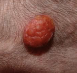
Histiocytomas can often be diagnosed by fine needle aspiration. The cells that comprise a histiocytoma have a characteristic microscopic appearance. Histiocytomas usually occur as a solitary, fast growing, non-painful, dome shaped lesion in the skin. Usually the surface is shiny and hairless, but with time they can become raw and ulcerated.
Although histiocytomas are classified as tumors, they are unique in that they are benign and often regress and disappear without treatment. Those that do not regress on their owner should be surgically removed and submitted for histopathology to confirm that they are indeed histiocytomas. There is another type of mass that is similar appearance to histiocytoma that can be more aggressive or malignant, therefore removal of any masses that are not resolving on their own is important. The prognosis for surgical removal of the histiocytoma is excellent.
The recommended treatment course is typically time and close monitoring. Surgery may be advised based on history, location, appearance and results of fine needle aspiration. It is important to maintain good communication until the growth completely regresses or is surgically removed.